

Surgical Conditions of the Upper Respiratory Tract
In this last installment in the series on equine respiratory conditions, we are going to look at disorders which are not caused by either infection or allergy as in the previous two articles. Instead, these conditions result from flawed anatomy or function of the horse’s upper airway. These issues are not curable with medicines and this has led to the development of a dedicated field of equine upper respiratory surgery.
Like many areas of equine veterinary medicine, whole textbooks have been devoted to these surgical conditions, so we must stick to the basics in this article. Luckily, however, the principles involved are extremely simple and easy to understand, despite the complicated-sounding surgical terms.
Anatomy of Area
At the top of the horse’s windpipe is the larynx. This lies at the back of the throat and has many functions. It is anchored to the skull via a series of thin bones, so the windpipe actually hangs off the larynx, which hangs off the skull. The larynx is of course hollow, like the trachea, and is essentially the entrance through which the air first passes en route into the lungs. At the entrance to the larynx, there are two flat cartilages called the arytenoid cartilages. Think of these like patio doors which slide apart to open onto your garden. When the horse wants to breathe in, these cartilages open up and stay open. When the horse is swallowing food, they close over to stop that food going into the windpipe. The movement of these cartilages is controlled by muscles and nerves – one set for each side.
Laryngeal Paralysis
In the most common surgical condition of the equine respiratory system, the left sided cartilage becomes paralysed and hangs down into the opening of the windpipe. Due to the huge pressures generated by the horse breathing during exercise, this floppy cartilage can get sucked right across the opening of the larynx, all but blocking air flow completely. The degree of paralysis is hugely variable, but affected horses will experience a significant reduction in air flow to the lungs, which will cause decreased oxygen levels in the muscles and a reduction in athletic performance. The cause of this paralysis is known to be a failure of the nerve on that left side to stimulate the muscle to pull the cartilage, and once it has happened, it’s permanent. The condition is much more common in large horses, eg a 17 hand thoroughbred is highly likely to have some degree of Laryngeal Paralysis.
These horses are completely unaffected when at rest, or even at moderate exercise, as their oxygen requirement is perfectly achievable even on a restricted airway. It’s only at hard work that the problem becomes a factor in performance.
Diagnosis of Laryngeal Paralysis
When an affected horse is cantered, and they start to breathe heavily, (creating negative airway pressures which suck in the paralysed cartilage), a whistling noise is heard on every inward breath. This is the sound made by air passing over the semi-closed larynx and having to whirl around before it gets down into the windpipe. It usually sounds exactly like someone blowing briefly over the top of an open bottle neck. As a vet, when vetting a horse for purchase, this is a crucial part of the 5 Stage Examination. The horse is usually cantered in a circle, and we listen carefully for any whistling sound. The ability to pick up this little soft whistling sound underneath the snorting breath sounds as the horse thunders around in a circle is an acquired skill. I watched my boss perform dozens of vettings before I felt confident that I could pick up a horse that ‘made a noise’ at exercise.
The diagnosis is generally confirmed via endoscope, where a camera on a flexible scope is passed up the nose and the entrance to the larynx is visualized. If the horse’s nose is occluded for a few seconds so that he breathes a few deep breaths after uncovering, the ‘lazy’ arytenoid can be seen lying down into the laryngeal entrance. In recent years it has become possible to endoscope the horse while it exercises, either by using a treadmill, (think huge static running machine for horses), or by having the scope in place in the horse’s throat while on the gallops. The rider carries the battery and light source and the images are recorded for subsequent analysis by the vet team.
Surgical Treatment
Laryngeal Paralysis is a condition which has been recognized for well over 100 years. The first Veterinary Surgeon to perform surgery on these horses was Sir Frederick Hobday back in the early 1900s. His operation involved opening up the larynx from the underside and removing a fold of tissue which would then cause scar tissue to form thereby ‘sticking’ the left arytenoid cartilage back in a more open position. If anyone recognizes the phrase – ‘getting a horse Hobdayed’, this is where it comes from. The Royal Veterinary College in London recently posted a 100 year old video of this operation being performed in its early days. To view click here.
For most horses, the starting point for surgery is the Hobday procedure with the additional removal of a fold of tissue below the affected arytenoid cartilage. This is called the Aryepiglottic fold (AE fold). The AE fold has the potential to bulge into the airway opening during exercise, and its removal eliminates one more potential barrier to air flow. These two procedures are relatively straightforward for an experienced respiratory surgeon, and owners and trainers are often keen to have them performed on a decent horse affected by laryngeal paralysis in the hope they will bring some improvement.
If this surgery is not successful, or in more extreme cases of Laryngeal Paralysis, a much more involved procedure called a Tie-Back may be performed. In this operation, a permanent suture is placed between the paralysed arytenoid cartilage and the back of the larynx, pulling the cartilage up and out of the airflow – literally the arytenoid cartilage is ‘tied-back’ in an open position.
The success of these surgical procedures is variable and depends a lot on the skill and experience of the surgeon. Tie-back operations depend on placing the suture with the correct tension on the arytenoid cartilage and this tension being maintained while the tissues around the surgical site heal and scar tissue forms. As you can imagine this does not happen every time!
The top surgeons can be flown around the world to operate on the most expensive racehorses and their names are known across the veterinary profession – nice job if you can get it! The procedures are not without problems however, the main one being that some damage can be done to the normal protective function of the arytenoids to protect against food entry during swallowing. Some horses after a Tie-Back procedure cough a lot, as they must constantly expel food and saliva from their windpipe.
Soft Palate Displacement
The soft palate is a horizontal muscular shelf at the back of the throat which helps guide food into the oesophagus. In the horse it should lie underneath the larynx at all times, apart from the few seconds when the horse swallows food, when the palate flips up to guide food away from the airway and into the gullet. When the palate is covering the larynx for this brief time, the horse can’t breathe (which is normal for all animals while swallowing). In some horses under hard exercise however, the palate can flip up for a second or two ‘by mistake’. This immediately cuts off the horse’s air supply and has a disastrous effect on its performance. A characteristic ‘gurgling’ noise is heard by the rider (usually a jockey) at this time.
Procedures which attempt to give the soft palate a stiffer, less ‘flappy’ edge, which is therefore less likely to flip above the larynx, are popular. This is predominately done via electrocautery, where high temperatures sear and stiffen the portion of the palate in contact with the larynx. A new technique where an injectable stiffener called Gentipin is used, seems to be promising.
An extensive surgical procedure where the larynx is pulled forward to ‘trap’ the palate in the correct position is performed in cases where other techniques have failed to satisfactorily correct the problem. Called the ‘Tie-Forward’ it involves the placing of permanent sutures to pull the larynx forward in the throat, hopefully trapping the palate in a permanent ‘underneath’ position.
Summary
Respiratory surgery in equine athletes, particularly racehorses, is a veterinary industry all of its own. While these surgical procedures undoubtedly improve the performance of many horses, they are by no means a complete solution in all cases. Many horses will fall out of racing because their ‘wind’ problems were too severe, or the surgical procedures just didn’t seem to work – often for no discernable reason. Happily though, these horses will be perfectly healthy and capable of becoming riding horses, hunters or eventers (maybe just not Olympic standard).
John Henderson B.V.M&S, M.R.C.V.S.
For support with good respiratory health view our recommended products:
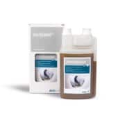
BRONCHOPULMIN 1L
Essential oils and Vitamin C to support respiratory health.
Bronchopulmin is a unique liquid feed supplement designed to restore and maintain a healthy and effective respiratory system and to support the immune system. It contains a blend of Essential Oils, such as Cineol and Eucalyptus oil, specifically chosen for their ability to support the respiratory immune system and soothe irritation. Additionally Thyme, Fennel and Black Cumin oil, support Bronchodilation. For more on derbymed’s Bronchopulmin click here.
 RESPIMIN 800G
Ultimate respiratory health supplement
Respimin is a feed supplement designed to support the equine respiratory tract. It is used to support horses diagnosed with respiratory conditions, and recovering from respiratory diseases such as acute, sub-acute and chronic infections (bronchitis, bronchiolitis and bronchopneumonia), viral respiratory diseases, respiratory allergies, and Recurrent Airway Obstruction (RAO). Its unique formulation encourages the release of mucous while reducing excessive coughing. For more on ARKequine’s Respimin click here.


